Microtissue model as a basis for predicting therapeutic outcomes of cartilage repair based on autologous cells
An autologous transplantation has the fewest side effects, but this approach does not always produce functional cartilage. A challenge in cell-based cartilage regeneration therapies is the identification of a ‘‘personalized diagnostic tool’’ to predict the chondrogenic potency of cells from patients who are going to be treated with autologous cells. Using our three-dimensional (3D) cell culture technique we could show clear differences in chondrogenic potential among individual donors whereas cells in 2D culture exhibited an identical chondrocyte profile. Therefore, our microtissue model might be the basis for an in vitro platform to predict the therapeutic outcome of autologous cell-based cartilage repair and/or a suitable tool to identify early biomarkers to classify the patients (Martin et al., 2017, Featured Article, Experimental Biology and Medicine).
In vitro tissue model for Experimental Pharmacology
In order to test the effect of drugs on joint cartilage, work is currently underway on the development of an in vitro tissue model, since the cartilage-typical physiology results primarily from the tissue formation with its typical extracellular matrix. A particular challenge in this project is the availability of cartilage cell lines that can be cultivated and propagated over a longer period of time with constant quality. To this end, methods for extending the lifespan and cell division as well as the generation of induced pluripotent stem cells (iPS) are used to develop cartilage cell lines. If required, these are then available at any time for the engineering of microtissues.
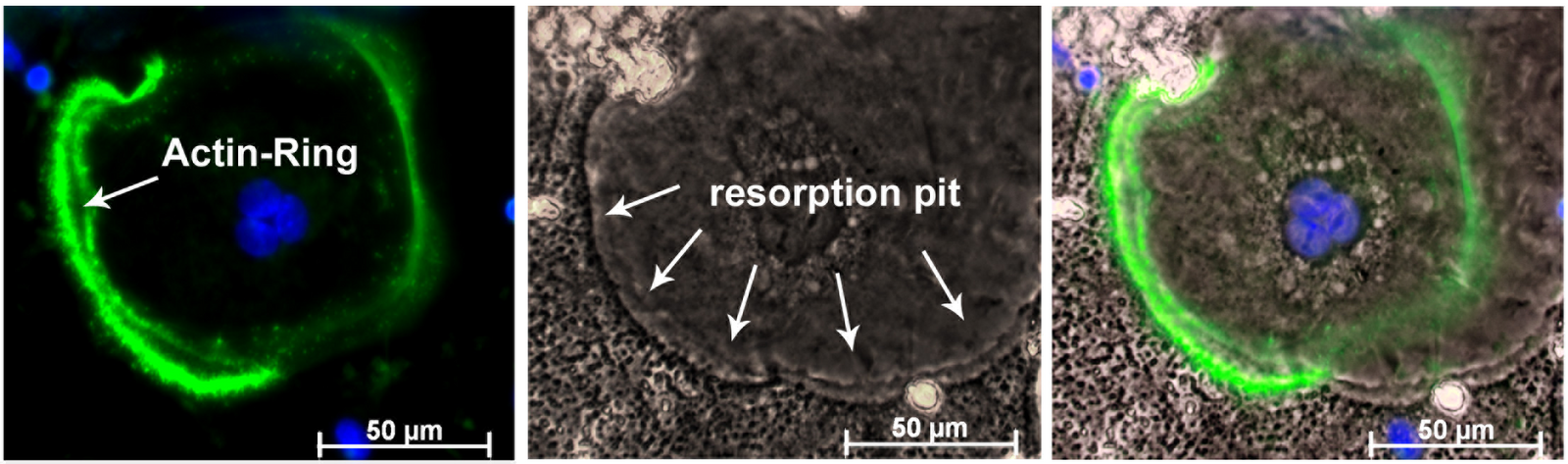
Investigation and diagnosis of osteoclast-specific diseases
Better knowledge of skeletal cell biology is the basis for understanding bone-specific diseases, such as osteoporosis, arthritis, and genetic conditions that affect bone growth and bone aging. Dr. Lutter studies how the skeletal cells function, how they communicate and regulate one another, with focus on the bone-resorbing osteoclast. Osteoclasts are large, mobile cells that begin their life as mononuclear hematopoietic cells. Under the influence of specific growth factors (MCSF and RANKL), the precursors can migrate to the site of bone resorption, attach tightly to the bone and secrete bone dissolving factors. To study how osteoclasts behave in natural habitat we developed an approach to assemble a bone-like extracellular matrix derived by human osteoblasts (ODEM, Lutter et al., 2010 Journal of Cellular Biochemistry). This special experimental setup enables our group to connect bone and cartilage research in a novel way.
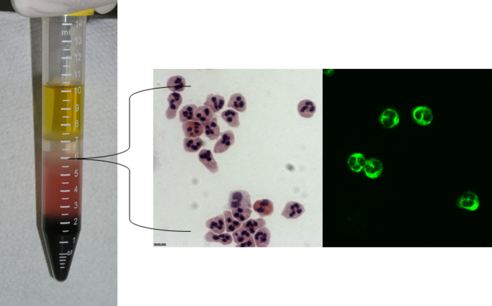
Cell-based test systems for the diagnosis of autoimmune diseases
In autoimmune diseases antibodies of a person’s own immune system are directed against the body's own structures. These diseases include e.g. type 1 diabetes and multiple sclerosis.A characteristic feature of autoimmune diseases is the formation of disease-specific autoantibodies (AAK) that are used as diagnostic markers. These autoantibodies are detected, for example, by fluorescence-based labelling of the appropriate antigen structures in cells. Special challenges in this process are suitable, well-characterized cell lines or freshly isolated cells, a preparation technique of the cells that guarantees the most comprehensive detection of the possible target structures of the AAKs, as well as an automated evaluation of the test system. These projects are carried out in close cooperation with Generic Assays GmbH in Dahlewitz.
Development of a novel test system for the autoimmune diagnostics of diseases of the nervous system
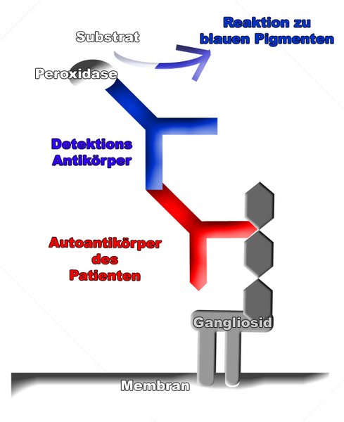
The project "Development of a novel test system for the autoimmune diagnostics of diseases of the nervous system" resulted in a direct transfer of knowledge and technology from the university to the diagnostics market.
The partners from the university (Prof. Ursula Anders) and cooperating companies - Generic Assays GmbH (Prof. Dirk Roggenbuck) and Attomol GmbH (Dr. Werner Lehmann) - worked together on an application-oriented research project. A test prototype was developed for the determination of autoantibodies against ganglioside antigens. Following the project period, clinical evaluation was carried out to determine the general quality parameters.
In this project, four biotechnology students were actively involved with their project work (sixth semester) and one student was actively involved during his bachelor's thesis. After completing his bachelor thesis, Mr. Milius was recruited by the Attomol GmbH as an employee and still works there today.
At present, the developed test system is manufactured by Attomol GmbH, which has created the technical and technological conditions for this purpose. Generic Assays GmbH successfully markets the product on the diagnostics market. The cooperative development and transfer work for this test system was honored with the Technology Transfer Prize 2005 of the Technology Foundation Brandenburg.