Chromatography and UV-Spectroscopy
Investigation of leaf pigments
Chromatography is the collective term for a family of laboratory techniques for the separation of mixtures. It involves passing a mixture dissolved in a "mobile phase" through a stationary phase, which separates the analyte to be measured from other molecules in the mixture and allows it to be isolated (Wikipedia).
Substances, which are able to absorb photons and therefore appear coloured, are called pigments. Using spectrophotometry (UV-VIS), the absorption of light by pigments can be measured at different wavelengths. The absorption signal versus irradiated wavelength is called absorption spectrum. The absorption maxima inform about the characteristics of these pigments. They can be consulted to characterize and quantify substances.
Liquid-liquid extraction of carotinoid and chlorophyll
Materials
- UV/VIS Spectrometer
- Laboratory centrifuge
- Water-bath
- Plant leafs
- Beakers, reactions flasks, reaction vessels, Pasteur pipette
- 90% methanol solution (Highly flammable, toxic; R: 11-23/25; S: 7-16-24-45)
- Petroleum benzine (Highly flammable, harmful; R: 11-52/53-65; S: 9-16-23-24-33-62)
Execution
- Boil 1-2g of leafs in a 90% methanol solution (approx. 20mL) using a water-bath
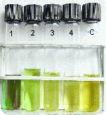
- Pour 5 mL of the extract with the same volume of petroleum benzine in a sealable test tube
- Shake it
- After the separation of the two liquid phases (which colours ?), remove the top phase (petroleum benzine) with a Pasteur pipette, and keep it for further analysis.
- Repeat three times the same process with petroleum benzine, and each time keep the top phase for spectrum analysis.
- Centrifuge the bottom phases (methanol)
- Dilute all samples by 1/10
Record spectrums between 350 and 750 nm for the original solution and for each of the separated phases. Which spectrums could you recognize?
Separation of leaf pigments (Liquid-solid extraction)
Materials
- UV lamp
- Plant leafs
- Water-bath
- Board chalk
- Methanol (Highly flammable, toxic; R: 11-23/25; S: 7-16-24-45)
- Petroleum benzine (Highly flammable, harmful; R: 11-52/53-65; S: 9-16-23-24-33-62)
Execution
- Dry pieces of chalk for 1 hour at approx. 100 °C
- Boil plant material in 15 ml methanol (water-bath), until a clear, highly concentrated solution appears
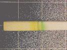
- Place the chalk vertically into a 25 mL beaker filled with chilled methanol extract
- When the solution rises approximately 10 mm high, place the chalk vertically in a 100 mL beaker filled with 5 mm petroleum benzine
- Pursue the development of the chromatogram till 1 cm from the top, and draw the front limit
- Place the chalk under the UV lamp
Fluorescent substances can absorb energy from ultraviolet radiation and immediately re-emit the energy as visible light. Observe the different colours.
Spectrometric analysis of chlorophyll A and chlorophyll B
In pigment mixtures the spectra of all individual components overlay and generate a combined spectrum.
If the Beer-Lambert law is valid: Etotal = al1.c1 + al2c2 + alncn
If the extinction coefficients of the individual pigments are known, their concentrations can be calculated from the total absorption spectrum. What’s more, Etotal is measured with n wavelength values. Each measured point is a function of two unknown variables which can be resolved.
For the two-component solution, chlorophyll A and chlorophyll B, the two red zones of the pigments are suitable for the differentiating measurement (664/647 nm in 80 % acetone) with the following extinction coefficients:
| l (nm) | Chl a (cm2 mg-1) | Chl b (cm2 mg-1) |
|---|---|---|
| 664 | 89.0 | 10.2 |
| 647 | 21.2 | 52.3 |
For our sample:
E664 = 89.0 CChl a + 10.2 CChl b
E647 = 21.2 CChl a + 52.3 CChl b
CChl a = 11.78 E664 – 2.29 E647 (mg/L)
CChl b = 20.05 E647 – 4.77 E664 (mg/L)
Extraction and measurement of the pigments must be operated very carefully, because the measurement taking place at 647 nm correspond to a steep absorption flank. The l-values must be recorded as exactly as possible with a spectral bandwidth less than 3 nm. The samples solutions must be turbidity free.
Material
- PE Lambda
- Mortar
- Glass filter and paper filter
- 100 mL measuring flask
- Sand
- Leafs
- Extraction solution: 80% acetone (Highly flammable; R: 11; S; 9-16-23.2-33) + 20% of an ammoniac solution (0,05M/L) (Corrosive, irritant; R; 34-37; S: 7-26-36/37/39-45)
Execution
- 1 g of leafs is weighed, cut roughly and finely grounded in a mortar with a pinch of sand and 3 mL of the extraction solution
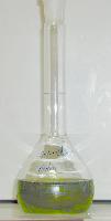
- Filter the homogenat with 2 paper filters and collect the extract in a 100 mL measuring flask. Fill up to 100 mL. The solution must be completely clear (without any particles)!
- Record a spectrum of the extract in a 1 cm cuvette between 350 and 760 nm
- (E750 value would be subtracted from the E647 and E664 values in the case it would be different from 0. This process would correct for eventual suspended particles).
Calculate with the corrected values (if necessary) the concentrations of chlorophyll A and B.
How is the relation between chlorophyll A and chlorophyll B?


