Presentation methods
With the SEM it is possible to visualize the objects to be examined in different ways. There are 3 detectors available on our instrument:
- Secondary electron detector (SE)
- Backscattering electron detector (BSE)
- Energy dispersive X-ray fluorescence analysis system (EDX)
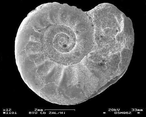
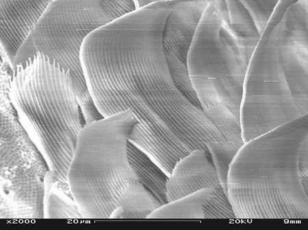
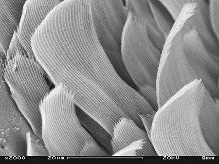
Depending on the use of the detector and the sample preparation, different aspects of the object to be examined are obtained. The following pictures show this on an ammonite (Black Jurassic, locality Belzmühle near Bielefeld), which was vaporized with coal after mounting:
SE detector
The side-mounted secondary electron detector provides an image of the surface topography of the specimen. In front of the scintillator is a grid-shaped collector, which is brought to positive voltage for secondary electron detection and thus forms a suction field for secondary electrons around the specimen, so that secondary electrons from parts of the specimen located away from the scintillator can also be registered by it.
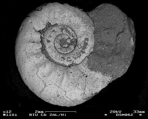
SE detector/BSE mode
When the collector is negatively biased, the secondary electrons are kept away from the scintillator and only the higher-energy backscattered electrons are registered, thus obtaining a backscattered electron image of the specimen surface. Since no extraction field is generated, the surface image exhibits a strong light/shadow effect.
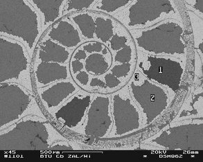
BSE detector
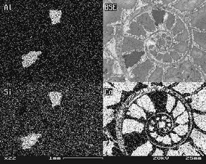
In contrast to the SE detector, the BSE detector symmetrically mounted under the objective with a YAP single crystal scintillator provides a depth image of the sample. The number of backscattered electrons depends on the density or the average atomic number of the respective structure (element contrast). The element contrast is superimposed by the specimen surface. In order to avoid this phenomenon, specimens are often examined in microsection. In the case of the ammonite, two areas with different element contrast can be seen in the image.
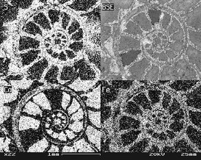
EDX detector
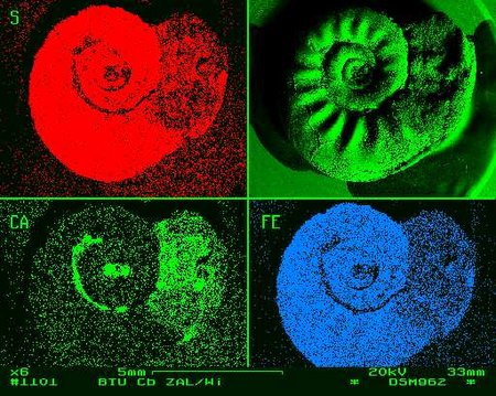
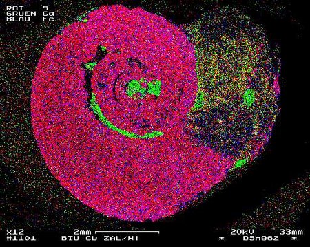
The energy dispersive X-ray fluorescence detector registers the X-ray quanta triggered by electron bombardment between up to 20kV. In addition to the non-specific bremsstrahlung, element-specific X-ray quanta are also registered, which can be used for qualitative (and quantitative see below) analysis of the elements up to now from sodium. In the meantime also the light elements starting from beryllium can be detected. Projection of the specific X-ray fluorescence onto the grid results in element distribution images with either single channels or common projection.
To use the EDX detector for quantitative analyses, the object must be flat so that the angle between the incident electron beam and the detector is constant. For this purpose, the object can be ground directly - as shown in the example of ammonite - or the material to be examined (e.g. soil, sand, biological material) is embedded in synthetic resin before grinding. The element contrast image (BSE detector) is examined. The quantitative analysis is performed after standardization with known samples or - much simpler - standardless after calibration with cobalt.
Filigree, non-self-conducting specimens, such as arthropods or specimens dusted on conductive foil, often show strong charging phenomena that prevent secondary electrons from being emitted "normally"; filamentous specimens may also move by repulsion due to charging phenomena in the microscopic image. The usual approach to such problems is to reduce the accelerating voltage of the electrons below 5 kV. Often samples that have not been made superficially conductive by vapor deposition or sputtering can be examined in this way. The disadvantage of this method is the significantly reduced resolution, which leads to clearly blurred images even at medium magnifications of 1000x or more. The BSE detector provides a remedy! The BSE signal is usually not influenced by charging phenomena! Even at medium and high magnification, detailed images can be obtained with high accelerating voltage of the electrons.
Copyright: All graphics are the property of ZAL and may not be republished without permission.Photos: Dr. Wolfgang Wiehe

