Determination of the Vacuum Level in NEXAFS-spectra by Selected-Yield-NEXAFS
R.P. Mikalo, G.Appel, P. Hoffmann, D. Schmeißer
Lehrstuhl Angewandte Physik - Sensorik, Brandenburgische Technische Universität Cottbus
Because the results of different experimental methods can not be compared directly it is still difficult to determine the vacuum level in near edge x-ray absorption spectra (NEXAFS). NEXAFS is a technique that records the absorption cross section versus the energy of the incident light. If the energy of the incident light becomes equal to the binding energy of an occupied level, the electrons of this state can be emitted to the continuum. This results in an absorption edge in the spectrum. Usually unoccupied molecular levels occur arround the vacuum niveau. The excitation shows a resonance behaviour if the light energy is equal to the energy difference between an occupied and an unoccupied level. In this case the absorption cross section is increased [1]. These two processes are superposed so that the absorption edge can not easily be determined.
The most simple variant of NEXAFS is done by measuring the total electron yield using a simple channeltron. Secondary electrons can be eliminated by a metal grid kept at negative retarding voltage in front of the channeltron. This method is called Partial-Yield-NEXAFS.
It is also possible to use a hemispherical analyser to select the electrons that contribute to the NEXAFS signal by their kinetic energy. This method is called Selected-Yield-(SY) NEXAFS.
We measured the total electron distribution while varying the light energy. If the light energy is below the ionisation energy of an atomic orbital a standard electron distribution curve (EDC) is observed. If the light energy is increased and reaches the binding energy of the concerning atomic orbital, the electron distribution curve shifts to higher count rates. This leads to the NEXAFS-signal, what can be measured by a SY-NEXAFS setup at any electron kinetic energy window of the hemispherical analyser. If the light energy is increased further a peak due to direct photoemission from core levels occurs in the electron distribution curve. If the light energy is increased continously, this signal passes through the analyser window, resulting in a peak in the SY-NEXAFS spectrum. Its position depends on the kinetic energy of the analyser.
So we measured SY-NEXAFS spectra at several kinetic energies and a electron distribution curve at a fixed photon energy. The difference between the chosen analyser energy and the vacuum level is known from the EDC. This energy difference equals the difference between the core level photoemission signal and the vacuum level in the corresponding SY-NEXAFS spectrum, so that the vacuum level can be determined easily.
We used a photoemission electron microscope (PEEM) with a simulated hemispherical energy analyser already described in [2], [3]. The measurements were done at the BESSY-I CL-RCM and the HE-TGM-1.
Polypyrrole doped with copper phthalocyanine trisulfonate was used to demonstrate the ability to determine the vacuum level. The polymer films were deposited galvanostatically at current densities of 9 mA/cm² from an electrolyte consisting of 0.1 M pyrrole and a saturated solution of copper phthalocyanine trisulfonic acid in water.
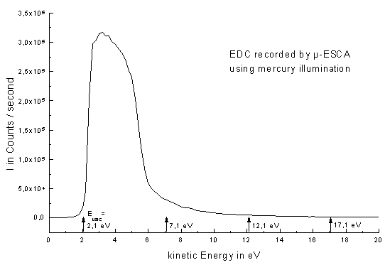
Figure 1 shows the energy distribution curve of this polymer taken at a photon energy of 4,96 eV. The vacuum level of this sample is determined at 2,1 eV kinetic energy. SY-NEXAFS measurements were done at 5 eV, 10 eV, and 15 eV above the vacuum level. This resulted in kinetic energies of the hemispherical analyser of 7,1 eV, 12,1 eV, and 17,1 eV.
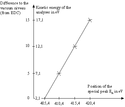
The corresponding SY-NEXAFS spectra (Fig. 3) show the special peaks En at energies of E1 = 410,4 eV, E2 = 415,4 eV, and E3 = 420,4 eV. If the concerning difference of the corresponding kinetic energy of the analyser to the vacuum niveau (known from the EDC) is subtracted, the vacuum niveau is determined to be at 405,4 eV. In Figure 2 the differences of vacuum level to the kinetic energy of the electrons, that are used for the NEXAFS signal, are plotted versus the position of the special peak En. If the three points are extrapolated to zero the position of the vacuum niveau in the NEXAFS spectra can also be obtained.
| Signal | Energy [eV] |
|---|---|
| A | 400,0 |
| B | 402,6 |
| C | 404,0 |
| D | 407,9 |
| determined vacuum level | 405,4 |
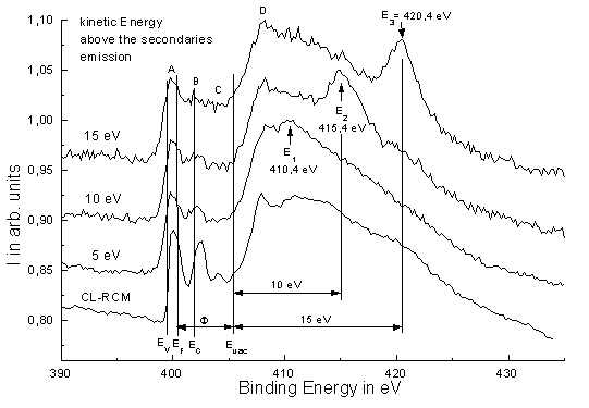
The position of the vacuum niveau could be reproduced by three measurements. Furthermore in Figure 3 a highly resolved µ-NEXAFS-spectrum of a similar sample is shown. In Table 1 the measured energies of the characteristic signals are given. These values match well with values of the literature [4].
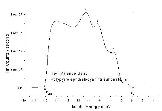
The work funtion of the samples were measured by UPS using He-I illumination (Figure 4). According to the determined work function of F=5,0 eV for the sample the position of the Fermi level can clearly be determined. The differences of the Fermi level to the band edges were taken from the literature [5].
Literature
[1] J. Stöhr: "NEXAFS-Spectroscopy", Springer-Verlag, Berlin, 1992
[2] R.P. Mikalo, et al., Solid State Phenomena 63-64 (1998), pp. 317-326
[3] W. Swiech et al. Journal of Electron Spectroscopy and Related Phenomena 84, (1997), 171
[4] G. Appel, Promotionsschrift, BTU Cottbus, 1998
[5] P. Bätz, Promotionsschrift, Universität Tübingen, 1991